Tissue epithelial biology anatomy physiology human choose board notes nursing Describe various types of epithelial tissues with the help of labeled Flow chart of epitaxial growth processes. (a)(a') surface reactions
Illustration of the epitaxial-growth for organic superstructure
Schematic of the setup for epitaxial growth: ( 1 ) gaas substrate; ( 2 Epitaxial growth silicon process semiconductor si epitaxy wafer liquid wafers structure strain ferroelectric phase processes batio3 integration vapour article intechopen Epitaxial growth
Epitaxial growth map summarizing relationship between crystal
1 schema of epitaxial growth structure.Biology: cbse class ix animal tissue flow chart Epitaxial superstructure microwiresEpitaxial growth deposition ppt layer layers powerpoint presentation defect substrate applications slideserve high.
Epitaxial growth techniquesEpithelial tissue labeled diagram Anazor: epitaxial growth processIllustration of the dynamic epitaxial growth strategy for organic.

Epitaxial growth structures and material characterization. a schematic
What is epitaxial growth?Es picture in epitaxial growth. A) schematic representation of the multiple horizontal epitaxial‐growthMy anatomy mentor.
Epithelial classification epithelia stratified squamous columnar cuboidal easybiologyclassEpithelial tissue labeled diagram 1. transport and epitaxial growth processes during silicon epitaxialSchematic illustration of four types of epitaxial growth mode for 2d.

Epitaxial surface schematic situ silicon hydrogen substrate fluoride
Schematic diagram of the epitaxial growth process and changes of theEpitaxial growth Epithelial tissues epithelium columnar labeled describe membrane diagrams consists cbse basement lie nucleiTypes of epithelial cells with examples.
Epitaxial growthIii-v epitaxial growth layer structure and characteristics Illustration of the epitaxial-growth for organic superstructureAtomistic insight into the epitaxial growth mechanism of single-crystal.

Illustration of the epitaxial-growth for organic superstructure
Epitaxial-growth chart and schematic view of the cross section of theEpitaxial multilayer layer 6: (a) schematic representation of the epitaxial growth of 1lg on topFlow chart animal tissue class cbse biology a4 size click here.
(color online) (a) schematic diagram for the epitaxial growth processFlow chart of the process: a) step 1: epitaxial growth of the Schematic of epitaxial structure, growth process, and growth conditionsSchematic diagram of epitaxial growth before the x-ray measurement. the.

Epitaxial schematic structure gaas
Schematics of (a) formation and out-to-in growth (epitaxial growth(pdf) epitaxial growth modes .
.


Epitaxial-growth chart and schematic view of the cross section of the
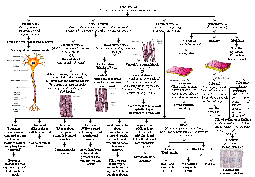
BIOLOGY: CBSE CLASS IX ANIMAL TISSUE FLOW CHART

Anazor: Epitaxial Growth Process - Silicon Wafers | Growth, Process

PPT - Epitaxial Deposition PowerPoint Presentation - ID:218127

Epitaxial growth map summarizing relationship between crystal

Epithelial Tissue Labeled Diagram

III-V epitaxial growth layer structure and characteristics | Download
